Uterine prolapse
Keywords: prolapse, uterus, ovine, eweA three-year-old Charollais ewe was presented approximately 24 hours after giving birth to twin lambs. No assistance was given during lambing. Serum ionized calcium concentrations were normal. This entry illustrates the typical approach to such cases in our clinic.
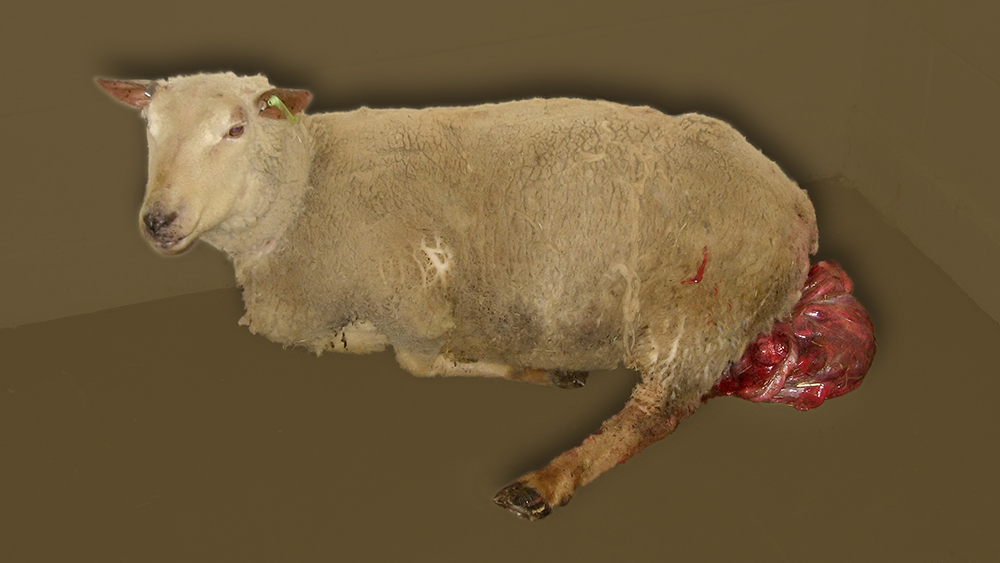
Image size: Click to enlarge to 1000 x 565 px
The uterus was inspected for damage and any fetal placenta that could be easily removed was removed. Because all the zoonotic threat of toxoplasmosis, Q fever and campylobacteriosis, gloves and masks (as seen here) should be worn.

After an epidural of 2 ml 2% lidocaine at the lumbosacral space, the ewe became recumbent.

Image size: Click to enlarge to 650 x 488px
The uterus was compressed and gradually replaced in a conventional manner over a period of approximately 10 to 15 minutes. The author prefers to use a towel, wrapped around the uterus for a non-slip compression surface; no sugar or other hygroscopic material is used. The uterus was then replaced in situ.

A Buhner-type of suture was placed into the vulvar lips (below) using a curved postmortem needle because the bovine Buhner needle that was available would have been too traumatic to use. Umbilical tape is being used as the fastening material. Note that a small incision has been made both dorsally and ventrally of the vulva to facilitate passage of the needle.

To facilitate the passage of the needle around the vulva, the needle enters and exits these incisions as shown below. This is the second pass of the needle around the perimeter of the vulva lips.

Image size: Click to enlarge to 650 x488px

Image size: Click to enlarge to 650 x488px

Image size: Click to enlarge to 650 x488px

The process is summarized below:

The umbilical tape was tightened so that the effective vulva opening was two fingers wide. See below.

The umbilical tape was tied in a bow for easy removal by the owner and excess umbilical tape was removed.


Image size: Click to enlarge to 650 x488px
The ewe was given a vaccine containing tetanus toxoid (Tasvax 8™) and was given ketoprofen at a dose of 3 mg/ Kg for analgesia. Five international units of oxytocin was given to stimulate involution of the uterus. Treatment with 22,000 IU/Kg of procaine penicillin G i.m. was started. Given twice a day, it and was continued by the owner for 5 days. The ewe was discharged to the care of the owner and she was instructed to remove the Buhner suture after approximately 1 week.
Unlike a prolapse vagina, a prolapsed uterus is unlikely to recur after subsequent lambing. Therefore affected ewes are usually kept in the flock.