OVINE IMAGES

How to use these images


IMPORTANT: To see an enlarged image, click on any image you see in LORI. Then, RIGHT click on the enlarged image to save it at its full size.


Saturday, April 17, 2021

Placentome variation. Ovine


Ovine placentation: placentome variation

Key words: multiplex, placenta, sheep, ovine

Sheep usually have about 80 to 90 placentomes with little variation. Caruncles are already present in the non-pregnant uterus (even before birth) and form future attachment points for cotyledons. The combination of caruncles on the maternal side and cotyledons on the fetal side form joint structures known as placentomes. Caruncles determine the sites for cotyledons on the surface of the chorion. Caruncles are not usually concave at the time of conception but develop into concave structures in most cases as pregnancy progresses. In some cases however, flat or even convex caruncles develop. As discussed below, this may be influenced by stress, nutrition and multiple fetuses.  


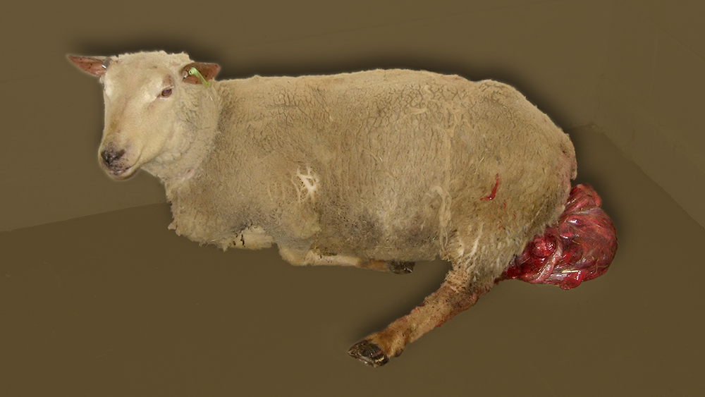
Figure 1. An image of a normal fetal-placental unit from a singlet pregnancy towards the end of gestation. Image size:1000 x 750px  


Figure 2. A cotyledon being withdrawn from a caruncle in this pregnancy. Although the majority of caruncles were concave in this pregnancy, a few caruncles (not shown) were indeed convex as occurs in cattle. Viewing the image at its highest resolution (right click on image) reveals the fine structure of the fetal villi. Image size: 4764 x 3456px


Figure 3. An area of caruncles with the cotyledons removed in most cases. As demonstrated by the small caruncle close to the center of the image, there can be considerable variation in their size. Image size: 4800 x 3456px

In times of nutritional stress, fewer placentomes are formed than otherwise. This has been shown in controlled studies on stressed and energy-deprived ewes. Interestingly, this not only affects the number of placentomes but their shape as well. In such states, some placentomes became flatter and even become bovine-like as alluded to above. Interestingly, these changes also occur in apparently normal pregnancies. Therefore one should exercise caution in studies of this nature. It not clear if caruncles established as concave can change their shape as pregnancy progresses but some data do suggest this may occur. i.e. 

Alexander G. (1963) reported that "....there was a marked tendency towards cotyledonary eversion; in nine out of ten placentae at both the 95th and 114th day the maternal tissue almost completely enveloped the foetal cotyledon, but at 135 days, the foetal tissue tended to surround the maternal in eight out of fourteen placentae, much as in the normal bovine cotyledon illustrated by Andersen (1926)."  

From these data and those reviewed by Vatnick et al, 1991, the author has a drawn a representation of  this situation:

Figure 4. The marked tendency towards cotyledonary eversion seen in some ovine pregnancies. These placentomes have been classified as types A to D. Type D is the most bovine-like of these. The green arrow questions whether this transformation occurs as pregnancy advances or if placentomes develop this architecture from the outset in some pregnancies. Interestingly, using a mathematical model, type D placentomes appear to the most efficient of all these forms of placentation. Using similar modeling, it has also been determined that ruminant placentation is highly efficient; 5 to 10 times more so than the diffuse, micro-cotyledonary placentation found in mares. Image size: 800 x 780px

This author has often wondered how essential the inter-placentomal areas are in maternal-fetal exchange in ruminants. As reviewed by Wooding and Burton (an excellent resource on placentation) there are small areolae over the mouths of the uterine glands in these areas but other than that, very little fetal-maternal exchange occurs in those areas. In fact, the inter-placentomal areas account for less than 5% of the total exchange at the end of ruminant gestation. 


Figure 5: An ovine pregnancy approximately 60 days duration showing some caruncles with a bovine (type D) appearance while others are clearly concave developing into the most common form of placentomes (type A). Right click to read labels and see in full size. Although the dark black areas on the endometrium are labeled as "Degenerate caruncles", this author is not at all certain if that is correct. Image size: 2599 x 1572px

Selected references:

Alexander G. 1963 Studies on the placenta of sheep J. Reprod Fert. 7:289-305

Vatnick I. et al. 1991. Growth and metabolism of the placenta after fetectomy in twin pregnant ewes. J. Development Physiology 15: 351-356

Vonnahme, K. A.  et al. 2006 Placentomal differentiation may compensate for maternal nutrient restriction in ewes adapted to harsh range conditions J. Anim Sci 84:3451-3459

Wooding, P. and Burton, G. 2008 in Comparative Placentation: Structures, Functions and Evolution. Pub. Springer eISBN: 9783540787976


Wednesday, March 3, 2021

Uterine prolapse

Uterine prolapse

Keywords: prolapse, uterus, ovine, ewe

A three-year-old Charollais ewe was presented approximately 24 hours after giving birth to twin lambs. No assistance was given during lambing. Serum ionized calcium concentrations were normal. This entry illustrates the typical approach to such cases in our clinic.


Image size: Click to enlarge to 1000 x 565 px

The uterus was inspected for damage and any fetal placenta that could be easily removed was removed. Because all the zoonotic threat of toxoplasmosis, Q fever and campylobacteriosis, gloves and masks (as seen here) should be worn.


Image size: Click to enlarge to 400 x 534px

After an epidural of 2 ml 2% lidocaine at the lumbosacral space, the ewe became recumbent.


Image size: Click to enlarge to 650 x 488px

The uterus was compressed and gradually replaced in a conventional manner over a period of approximately 10 to 15 minutes. The author prefers to use a towel, wrapped around the uterus for a non-slip compression surface; no sugar or other hygroscopic material is used. The uterus was then replaced in situ.


Image size: Click to enlarge to 400 x 534px

A Buhner-type of suture was placed into the vulvar lips (below) using a curved postmortem needle because the bovine Buhner needle that was available would have been too traumatic to use. Umbilical tape is being used as the fastening material. Note that a small incision has been made both dorsally and ventrally of the vulva to facilitate passage of the needle.


Image size: Click to enlarge to 400 x 534px

To facilitate the passage of the needle around the vulva, the needle enters and exits these incisions as shown below. This is the second pass of the needle around the perimeter of the vulva lips.


Image size: Click to enlarge to 650 x488px

The process is summarized below:


Image size: Click to enlarge to 650 x488px

The umbilical tape was tightened so that the effective vulva opening was two fingers wide. See below.


Image size: Click to enlarge to 650 x488px

The umbilical tape was tied in a bow for easy removal by the owner and excess umbilical tape was removed.

Image size: Click to enlarge to 650 x488px

The ewe was given a vaccine containing tetanus toxoid (Tasvax 8™) and was given ketoprofen at a dose of 3 mg/ Kg for analgesia. Five international units of oxytocin was given to stimulate involution of the uterus. Treatment with 22,000 IU/Kg of procaine penicillin G i.m. was started.  Given twice a day, it and was continued by the owner for 5 days. The ewe was discharged to the care of the owner and she was instructed to remove the Buhner suture after approximately 1 week.

Unlike a prolapse vagina, a prolapsed uterus is unlikely to recur after subsequent lambing. Therefore affected ewes are usually kept in the flock.


Saturday, January 30, 2021

Fetal genital tract and adnexa.

Keywords: ewe, ovine, lamb, uterus, term, ovaries

Uterine anatomy:


Figure 1. The genital tract of a Texel fetus (one of triplets) at approximately 130 days gestation. For scale, it is superimposed on the fetus itself in the upper image. Despite the presence of a male fetus in this pregnancy, mullerian development was normal in this female. For more on freemartinism in sheep, see another posting in LORI. Image size: 924 x 743 px

In a late gestation ruminant fetus, the author has noticed that the fetal uterus is usually tightly coiled as it is in adults during estrus. That appears to have been the case here too. This may be a response to elevated of placental estrogen secreted towards the end of gestation. Also note the dark fluid accumulation in the expanded infundibulum; reminiscent of hydropsalpinx. This has also been noted occasionally by the author in other ruminant fetal tracts. Its significant is unknown.

The reader is reminded that all the oocytes this fetus would ever have, were present in these ovaries at this time. Meiosis had indeed started but only progressed about half-way through meiosis 1 (reduction division) in those oocytes. The oocytes were still diploid (but had double the amount of DNA compared to somatic cells) when this photograph was taken. If this fetus had survived until puberty, an oocyte would be selected for ovulation (if it had not been wasted by regression during a diestrous follicle wave) and meiosis would resume under the effect of an LH surge during estrus. This division would form a polar body. Almost immediately, the mitotic phase of meiosis (meiosis 2) would follow and in theory, another two polar bodies would have been formed, stimulated by fertilization itself. In most vertebrates, fertilization is required to complete meiosis 2, in others not. Occasionally the first polar body does not divide, leaving only two polar bodies under the zona pellucida at the time of fertilization.

Recall that the mitotic phase of meiosis (meiosis 2) occurs almost immediately after the completion of reduction division. This occurs so quickly that there is not enough time for DNA to double. However, this is not a problem because DNA is doubled in oocytes just as meiosis starts, long before birth, in the early life of an embryo.  Again, all the oocytes in the fetal ovaries shown here had already doubled the amount of DNA they would have had at the time of ovulation.

Note: In males, there is no fixed number of gametes; diploid spermatogonia are continually replaced along the basement membrane of seminiferous tubules. Also, males do not experience LH surges. Instead, baseline LH secretion constantly stimulates spermatogenesis.

Ovarian function:

It is interesting to re-visit the quandary of why it is that corpora lutea are present in the ovaries at term, yet ewes can be ovariectomized after 60 days of gestation with no effect on lamb birth weight and survival. The ovaries of the ewe in this case were examined and as expected, at least three corpora lutea were present in the ovaries. See figure 2.


Figure 2. Corpora lutea in the ovaries of a ewe at term. Image size: 800 x 544 px

Another valuable view of this pregnancy was to examine structures that contribute to umbilical anatomy; a theme common to all mammals. In the following image the reader can appreciate how fetal structures are replaced in situ by their equivalents in the adult.


Figure 3. Structures surrounding the intra-abdominal portion of the umbilical cord, showing how they are replace by their analogs in the adult animal. Image size: 986 X 517 px
  
Selected references:

Al-Gubory, K.H. et al. 1999. Effects of luteectomy on the maintenance of pregnancy, 
circulating progesterone concentrations and lambing performance in sheep. 
Reprod.Fertil.Dev. 11(6):317-22

Clift, D and Schuh, M. 2013 Restarting life: fertilization and the transition from meiosis to mitosis. Nat Rev Mol Cell Biol. 14:549-62

Cooper, G M. The Cell, 2nd edition. A Molecular Approach. Sinauer Associates; 2000.ISBN-10: 0-87893-106-6


Saturday, April 18, 2020

Ventral rupture and herniation.

Keywords: ventral hernia, ovine, ewe, premature

A primiparous Texel ewe presented for ventral rupture at approximately 144 days of gestation. Her udder was severely edematous and there was also considerable sub-cutaneous hemorrhage. She required help to walk from the truck to her stall. Her appetite was depressed but a a chemistry screen revealed that she was not ketotic. Her CBC and chemistry results were normal. Vitamin E and selenium supplementation in the 74 head flock were optimal. All common vaccinations had been given. The only recent change in management was the introduction of a large Suffolk ram.


Figure 1.  A ewe presented for ventral rupture in late gestation. Post mortem examination showed that the mesometrium had been ripped from the peritoneal surface creating a defect in the peritoneal wall (inset at lower left)  with the uterus come to lie beneath only a single muscle layer and the skin. Image size: 2788 x 1866 px

Although trauma and the weight of the uterus were suspected as inciting causes in this ewe, many observers have stated that ventral hernia may occur without obvious trauma. Rupture of the prepubic tendon is frequently mentioned in the context of ventral rupture in mares (although this is not necessarily accurate). In several cases, a similar assertion has been mentioned with regard to ewes. However, the author is not aware of any postmortem dissections that have confirmed that view.

Case management:

Because of an anticipated failure of the ewe to exert an abdominal press in second stage parturition, it was decided to deliver her lambs via cesarean section.To mature the lungs of her lambs in utero, 8 mg of dexamethasone was given to the ewe IM.  Due to a declining condition of the ewe and a limited budget, the elective C-section was planned for day 145, only one day after presentation. Optimally, more time (~36 to 48 hours) should be allowed for lung maturation after dexamethasone treatment.

Care of neonates:
An epidural anesthetic and inverted "L" were used for the C-section and two live lambs (figure 1) and one dead lamb were delivered. As per usual, suction was used to clear airways of the lambs. Oxygen was supplied using a face mask. Vigorous rubbing and ~ 20 mg of doxapram hydrochloride was used to stimulate respiration in the neonates. Each lamb was tube fed ~35 ml of colostrum (from another ewe on the farm) immediately after birth. When the lambs had become fully vitalized they were offered more colostrum; an essential source of energy as well as antibodies. The final volume consumed by each lamb was just over 100 ml each. The lambs were examined for sex, cleft palate and other abnormalities and their umbilical cords disinfected. In this case chlorhexidine solution was used. The farmer was reminded to repeat this in 24 hours. Until the owner fetched the lambs, they were held in a stall under an infrared lamp at ~ 36 degrees C.

Note: Current literature discourages the use of doxapram in neonatal resuscitation.  The use of  the GV26 acupuncture site in the philtrum should be considered instead.

The ewe was euthanized.

Postmortem examination:
At postmortem (see figure 1) there was evidence of rupture of the mesometrium and the peritoneal surface on the left-hand side. The uterus was separated from the skin by only a single, stretched muscle layer. It was concluded that an accident such butting, falling or jumping had caused the heavy uterus (containing large Suffolk x Texel triplets) to rip free of the peritoneal surface.

Although trauma and the weight of the uterus were suspected as inciting causes in this ewe, many observers have stated that ventral hernia may occur without obvious external trauma (butting etc.). Rupture of the prepubic tendon is frequently mentioned in the context of ventral rupture in mares (although this is not necessarily accurate). In several cases, a similar assertion has been mentioned with regard to ewes. However, the author is not aware of any postmortem dissections that have confirmed that view.


Selected references:

Huso, B. T. 1941 "Rupture of the Prepubic Tendon of a Ewe," Iowa State University Veterinarian: 3: Article 17

Mahdi A.S. 2015 Ventral abdominal and umbilical hernia in sheep in Babylon. Euphrates Journal of Agriculture Science. 7: 1-7

Monteiro G.A. et al. 2019 Prepubic tendon rupture in ewe. Vet.eZootec.25:085-088

Noakes. D.E., Parkinson T.J.and England G.C.W. Author and Eds. 2009. Arthur's veterinary reproduction and Obstetrics.  Saunders. Chapter 10 pages 241 to 242

Tirgari M.1980 Ventral hernia in the sheep. Vet Rec.106: 7-9.


Tuesday, April 14, 2020

Scrotal hematoma/seroma and scrotal abscessation

Case 1: Scrotal  hematoma

Keywords: ovine, ram, scrotum, hematoma, infertility, fertility

A Texel ram approximately 4 years of age was presented with a history of  weight loss and right scrotal enlargement of approximately 2 to 3 weeks duration. This enlargement was semi-firm and the ram resented palpation in this area. The owner had treated the ram with penicillin and trimethoprim-sulfa for five days before presentation.

Figure 1. Enlargement of the right half of the scrotum in a 4 year old Texel ram due to a hematoma. Aseptic tapping of the right side of the scrotum revealed clear serum. Image size: 1000 x 755px

Ultrasonography showed the presence of a large hematoma in the right side of the scrotum and an apparently normal testis in the left side of the scrotum. See figure 2.

Figure 2. A hematoma within the right half of the scrotum (lower insets). Fibrin clots had displaced the right testis dorso-medially making it difficult to image; only the hematoma and part of the seminiferous cord within the right half of the scrotum (lower right) were visible. Cross sections and longitudinal sections of the left testis (upper insets respectively) suggested that the left testis was normal. Image size: 1039 x 825 px

As shown in figure 1, aseptic tapping of the hematoma revealed the presence of serum, presumably the result of  blood clotting. The serum sample was smeared and stained; it was acellular and no bacteria were found. However, there was a substantial increase in the peripheral neutrophil count (approximately double normal values) but no other suggestion of septicemia. The animal was bright alert and responsive throughout its stay in the clinic therefore the origin of the neutrophilia remained an enigma.

Surprisingly, electro-ejaculation produced a semen sample that was normal in appearance, with excellent motility and morphology. The ram was discharged and the owner was told that the animal could be used for breeding as soon as required. The farmer was also advised of the risk of scrotal abcessation and the need to monitor the ram's scrotum and fertility.

It was surmised that this seroma was the result of trauma as several other rams were housed together with the ram.

Case 2: Scrotal abcessation. 

Keywords: scrotum, ram, ovine, abscess, peritoneum, omentum, infertility

A mature Texel ram of uncertain age was admitted with a a large abscess. This was substantiated by ultrasonography (figure 3)

Figure 3. Ultrasonography adjacent to the right testis showing pockets of fluid (presumed to be pus) in a fibrous mass surrounding the right testis. Image size: 767 x 484 px

Aseptic tapping of the the right half of the scrotum (figure 4) revealed large amounts of pus.

Figure 4.  Tapping of the enlarged mass over the right testis in a ram with an enlarged scrotum, revealing a mixture of blood and pus. Image size: 1200 x 931 px

The ram was euthanized and submitted for postmortem examination. There was inflammation and hemorrhage in the tunica dartos around the right testis. There was also substantial pus accumulation in the vaginal cavity, under the visceral surface of the parietal vaginal tunic, adjacent to  the right testis.  There were also accumulations of pus in abscesses around the spermatic cord. See figure 5.

Figure 5. Inflammation and edema within the right tunica dartos surrounding the right testis. The intact parietal vaginal tunic surrounding the right testis can be seen in the same image. In the image to  the right, the parietal vaginal  tunic has been incised, revealing a mass of interconnected abscesses. Image size: 1200 x 880 px

The abscesses and fibrous tissue formed a mass beneath the visceral tunic of the right testis had enlarged sufficiently to ablate most of the vaginal cavity around the right testis. The shallow remnant of the vaginal cavity viewed from the peritoneal cavity is shown in figure 6.

Figure 6. A shallow remnant of the right vaginal cavity. The vaginal cavity normally surround the entire testis and epididymus. Inflammation beneath the vaginal tunic resulted in inflammation that caused adhesions between the visceral and parietal vaginal surfaces, diminishing the capacity of the vaginal cavity. Image size: 2044 x 2504 px

In addition to the scrotal abscesses, numerous abscesses were found in the inguinal lymph nodes and mesentery (figure 7) suggesting that the scrotal abscesses were part of systemic disease.

Figure 7. One of many peritoneal abscesses present in a ram with peri-testis abscessation. Image size: 1267 x 1454 px

Budget constraints did not allow bacterial culturing. Although other bacteria are sometimes involved in scrotal abscessation (see the following case) literature suggests that Corynebacterium pseudotuberculosis was the likely pathogen in this case.

Case 3: Scrotal abcessation. 

Keywords: scrotum, ram, ovine, abscess, E.Coli, infertility

A 2 1/2 year old companion animal ram of mixed breed was presented for an enlarged scrotum. As seen in figure 8, the scrotum was so dependent that it dragged beneath the animal's legs, causing trauma and smearing the ground with blood.

Figure 8. A grossly enlarged scrotum in a mixed breed ram. Note how the scrotum was so heavy and dependent that it dragged on the ground. An outline of  the left testis is clearly visible. Image size: 1265 x 791 px

This ram had sired at least three lambs prior to the onset of the swelling which was first seen about nine months before presentation. There was no history of injury. The ram was eating normally and passing feces at the time of presentation but according to the owner, had been mildly obtund for the preceding three days. The rDVM had sampled the swollen mass by needle puncture, revealing pus. Culture of that pus revealed E. coli.

Ultrasonography of the scrotum revealed multiple pockets of fluid with small echogenic particles suggesting the presence of multiple abscesses in the scrotum. There was no evidence of intestinal contents in the scrotum and only the left testis could be seen on ultrasonography.

Figure 9. Ultrasonography of the right side of the scrotum of a mixed breed ram, showing two abscesses (of many). The left testis appeared to be normal. Image size: 798 486 px

Due to the value of the ram as a companion animal, it was not euthanized. Instead, the scrotum was removed under general anesthesia. Severe blood loss during surgery necessitated hospitalization for several days after removal of the scrotum.

The scrotum weighed 14 kg after removal:  The left testis appeared to be normal in size. An impression smear of the cut surface of that testis and its epididymis revealed the presence of spermatozoa. A histological section of the left testis also showed that spermatogenesis was progressing normally.

Numerous incisions into the right testicular mass revealed many compartmentalized abscesses.

Figure 10.  A collage of images showing the content of the scrotum of a mixed breed ram. All images are viewed from the cranial aspect of the scrotum. Image A shows the amputated scrotum. As in image B, all images show the presence of the left testis (large blue arrow) Image C shows pus flowing from an incision in the right side of the scrotum. Image D shows a collection of red arrows, indicating many of the interconnecting abscesses in the right side of the scrotum. The only possible remnants of the right testis are indicated by the black arrows. Image size: 1904 x 1615 px

As was the case for the rDVM, culture of the pus from one of these abscesses also revealed a pure culture of E. coli.

Although a thorough dissection of the right testicular mass was undertaken, no obvious remnant of the right testis seminiferous epithelium was seen. A small section of what may have been seminiferous epithelium was examined histologically. This section was difficult to interpret but the author believes that the remnants of seminiferous tubules could be seen, together with numerous necrotic inflammatory cells. Remarkably, spermatogenesis appeared to be progressing normally in the left testis despite its proximity to this inflammatory process.

Selected references:

Chhalgari M.U. 2017. Caseous lymphadenitis in small ruminants: An overview on reproductive implications. https://www.researchgate.net/publication/316582742

Lacasta D.I. et al. 2009. Unilateral scrotal pyocele in ram caused by Staphylococcus capitis. Aust Vet J. 87:484-486.

Williamson P. and Nairn M E. 1980. Lesions caused by Corynebacterium pseudotuberculosis in the scrotum of rams. Aust Vet J. 56:496-498.


Scrotal (inguinal) hernias 

Keywords: scrotum, ram, hernia, intestine, infertility

Note: In normal males, the testis is covered intimately by the tunica albuginea. In turn, those structures are contained within the visceral vaginal tunic. The visceral vaginal tunic on the left and right of the testis fuses caudal to the testis where the fused portion envelopes the epididymis and ductus deferens. The vaginal tunic then reflects parietally on either side of the testis, forming the vaginal cavity between the parietal and visceral layers of the tunic. The vaginal cavity is continuous with the peritoneal cavity but the vaginal canal is normally narrow enough to prevent viscera from entering the vaginal cavity.

Note on terminology: 
Hernias are named according the location in which they are located (hiatus, umbilical, abdominal, femoral etc) and whether or not the pass through tissue barriers that did not exist previously such as muscle and connective tissue walls (direct) or into cavities that did exist previously, such as the inguinal canal or congenital cavities around the umbilical cord (indirect).

The term inguinal hernia is commonly used to in human medicine. This is because inguinal hernias in men or boys are usually found in the inguinal canal. However, inguinal hernias in humans also progress to occupy both the scrotum as well as the inguinal canal, yet they do not assume the name of this new location. This is almost exclusively a tradition in human medicine. It is probably because inguinal hernias are usually noticeable in animals only once they have become scrotal in location. Therefore veterinarians refer to  them as scrotal hernias. To be completely accurate, perhaps one should refer to these as "inguino-scrotal" hernias.

We have used the term scrotal hernia in this entry. When the inguinal canal and vaginal cavity serve as the hernial sac (usually containing intestines) the condition is referred to as an indirect scrotal hernia.  In the case of direct scrotal hernias, the hernial sac enters the scrotum but comes to lie outside the vaginal cavity. Direct scrotal hernias are very rare rare in domestic animal. The cases described here are indirect hernias.

Case 1. Scrotal hernia in a mature Texel ram. 

This ram (precise age unknown) was presented for routine breeding soundness evaluation. Due to the presence of the hernia and the possibility of this being a heritable trait, the ram was culled from the flock.

Figure 1. A right-sided scrotal hernia in a mature Texel ram. A, B and C are ultrasounic cross sections of the scrotum at corresponding levels. Intestines are visible in the vaginal cavity at level A while level B shows only fluid and fibrin. The hernia had not yet progressed to level C. Therefore only cross sections of the left and right testes are seen in that ultrasonograph. Image size: 1624 x 1306 px

Case 2. Massive scrotal hernia in a mature ram.

These images (15 years old) show a massive unilateral, intestinal hernia in the scrotum of an infertile Texel ram. Its age and breed were not recorded in the author's image bank.

This condition is easily diagnosed by ultrasonography but that was not used in this case. The ram was euthanized and went directly for a post mortem examination.

Figure 2.  A massive scrotal hernia in a Texel ram where the left vaginal cavity was distended enormously by loops of intestine. Almost undoubtedly, both testes would have experienced an elevated environmental temperature, resulting in suppression of spermatogenesis and infertility. Image size: 1463 x 1179 px

In this ram, the inguinal canal became dilated to the point where intestines escaped into the left vaginal cavity; a classic indirect inguinal hernia. However, this case was remarkable because a large volume of serum and fibrin accumulated around the left testis, between its tunica albuginea and the visceral layer of the left vaginal tunic. Presumably, vascular embarrassment in the left inguinal canal caused plasma to escape from the pampiniform plexus and other vessels around the left testis. This then dissected into the potential space between the tunica albuginea over the left testis and the left visceral vaginal tunic. Eventually, large amounts of fibrin and serum accumulated in this space.

Perhaps obviously, this ram would have been infertile because of a temperature insult to the left testis caused by the presence of intestines, combined with insulation of the testis by the surrounding layer of serum and fibrin. Spermatogenesis may have been been present  in the right testis but its smaller-than-normal size suggested otherwise.

The heritability of scrotal hernias in rams is not well documented; some authors stating that it is heritable, other deferring to uncertainty.  Scrotal herniation is however, a well documented heritable condition in pigs. Therefore it seems advisable not to repair scrotal hernias in rams.  It is also possible however, that scrotal hernias may result from trauma, and especially in group-housed rams.

Selected references


Al-Sobayil F.A.and Ahmed A.F.2007.Surgical treatment for different forms of hernias in sheep and goats J Vet Sci. 8:185-191


Murray. R. M. 1969. Scrotal abnormalities in rams in tropical Queensland with particular reference to ovine Brucellosis and its control. Australian Vet.J. 45: 63-67

Roberts, S. J. (ed).1986. Veterinary obstetrics and genital diseases (Theriogenology). Published by author, Woodstock, VT 05091. ISBN-10: 9997670922 ISBN-13: 978-9997670922. pp 822-823.

Roberts, S. J. 1988. Scrotal hernia in rams. A case report. Cornell Vet. 78: 351-352.

Scott P. 2013. Ram breeding soundness – some common scrotal and testicular abnormalities. UK Vet Livestock Vol. 17, No. 4 https://www.magonlinelibrary.com/doi/abs/10.1111/j.2044-3870.2012.00126.x

Monday, June 5, 2017

Inguinal (scrotal) hernias

Keywords: ovine, ram, scrotum hernia

These illustrations are more than 30 years old but still of interest.


Figure 1.  A massive unilateral, inguinal hernia in the left side of the scrotum of a ram. Image size: 1000 x 806px

This ram was presented for investigation of long-standing enlargement of its scrotum. Due to the age of the animal and the potential for life-threatening developments, it was euthanized.

Various medical dictionaries define a hernia as "... a bulge or protrusion of an organ through the structure or muscle that usually contains it". This condition therefore, should be referred as an inguinal hernia rather than a scrotal hernia. This is based on the fact that the scrotum itself has not been penetrated. An analogy would be to refer (incorrectly) to a diaphragmatic hernia as a thoracic hernia. 

In the case of a direct hernia, the protruding organ exits through a torn muscle layer/s. In direct inguinal (scrotal) hernias, the hernia has no guiding path and therefore, seldom protrudes far into the scrotum. More commonly, inguinal hernias are indirect; described as such because they do not penetrate muscle layers. Instead, viscera enter the internal inguinal opening and occupy the vaginal cavity of the scrotum.  

The condition in this ram is correctly be referred to as an indirect inguinal hernia.  

In normal males, the testis is covered intimately by the tunica albuginea. In turn, those structures are contained within the visceral vaginal tunic. The visceral vaginal tunic on the left and right of the testis fuses caudal to the testis where the fused portion envelopes the epididymis and ductus deferens. The vaginal tunic then reflects parietally on either side of the testis, forming the vaginal cavity between the parietal and visceral layers of the tunic. The vaginal cavity is continuous with the peritoneal cavity but the vaginal canal is normally narrow enough to prevent viscera from entering the vaginal cavity.

In this ram, the inguinal canal became dilated to the point where intestines escaped into the left vaginal cavity; a classic indirect inguinal hernia. However, this case was remarkable because a large volume of serum and fibrin accumulated around the left testis, between its tunica albuginea and the visceral layer of the left vaginal tunic. Presumably, vascular embarrassment in the left inguinal canal caused plasma to escape from the pampiniform plexus and other vessels around the left testis. This then dissected into the potential space between the tunica albuginea over the left testis and the left visceral vaginal tunic. Eventually, large amounts of fibrin and serum accumulated in this space.

Perhaps obviously, this ram would have been infertile because of a temperature insult to the left testis caused by the presence of intestines, combined with insulation of the testis by the surrounding layer of serum and fibrin. Spermatogenesis may have been been present  in the right testis but its smaller-than-normal size suggested otherwise. Certainly, this ram would not be suitable for breeding.

A less dramatic scrotal hernial in a Texel ram is show in figure 2.


Figure 2 A scrotal hernial in a Texel ram. The typical ultrasound findings (showing intestinal loops within the scrotum) is shown within the inset. Image size: 1000 x 1334px

Inguinal hernias in rams are not common and inguinal herniation in rams has received little attention in the literature, especially in contrast to boars, bulls and stallions where the condition is considered heritable. There is however, some suggestion that inguinal hernias are also heritable in rams. In light of these statements it is surprising to find several reports of surgical repair of inguinal hernias in both sheep and goats.

Selected references:

Murray. R. M. 1969. Scrotal abnormalities in rams in tropical Queensland with particular reference to ovine Brucellosis and its control. Australian Vet.J. 45: 63-67

Roberts, S. J. 1988. Scrotal hernia in rams. A case report. Cornell Vet. 78: 351-352.

Roberts, S. J. (ed).1986. Veterinary obstetrics and genital diseases (Theriogenology). Published by author, Woodstock, VT 05091. ISBN-10: 9997670922 ISBN-13: 978-9997670922. pp 822-823.

Scott, P. 2012. Ram breeding soundness - some common scrotal and testicular abnormalities. Livestock. 17: 37-41